




Kidney Cancer/ Renal Cancer
A significant proportion of kidney cancers are discovered incidentally — not because a patient presented with symptoms, but because an ultrasound or CT scan was performed for an unrelated reason. This incidental detection is increasingly common and often results in earlier-stage diagnosis, which generally allows for more treatment options. Incidental detection often means earlier-stage disease, which frequently allows nephron-sparing surgical options and more favourable outcomes. When kidney cancer is found this way, further imaging with a CT scan or MRI is performed to characterise the lesion before treatment planning begins.
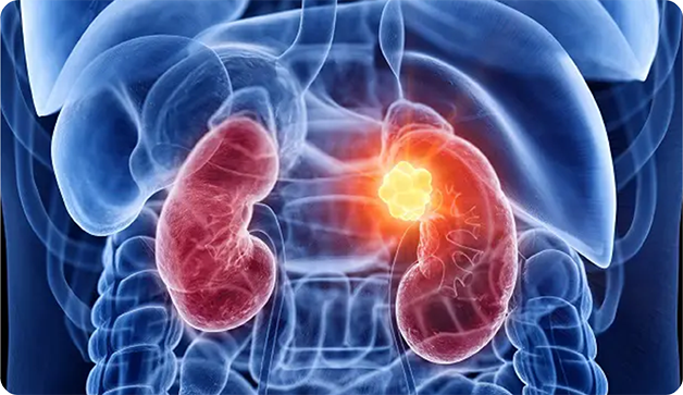


How Is Kidney Cancer Detected During Routine Scans?
A significant proportion of kidney cancers are discovered incidentally — not because a patient presented with symptoms, but because an ultrasound or CT scan was performed for an unrelated reason. This incidental detection is increasingly common and often results in earlier-stage diagnosis, which generally allows for more treatment options. Incidental detection often means earlier-stage disease, which frequently allows nephron-sparing surgical options and more favourable outcomes. When kidney cancer is found this way, further imaging with a CT scan or MRI is performed to characterise the lesion before treatment planning begins.
Types of Kidney Cancer
Kidney cancer encompasses several distinct subtypes, each with different biological behaviour, prognosis and sensitivity to treatment.
Clear Cell Renal Cell Carcinoma (ccRCC): The most common subtype, accounting for approximately 70–75% of all renal cell carcinomas. Characterised by cells with clear cytoplasm due to glycogen and lipid accumulation. Strongly associated with VHL gene mutations. Responds well to VEGF-targeted therapies (sunitinib, cabozantinib, axitinib) and immunotherapy combinations (nivolumab-ipilimumab, pembrolizumab-axitinib).
Papillary Renal Cell Carcinoma (pRCC): The second most common subtype (10–15%). Classified as Type 1 (less aggressive) and Type 2 (more aggressive). MET pathway alterations are common. May benefit from MET-targeted therapy in selected cases.
Chromophobe Renal Cell Carcinoma: Accounts for approximately 5% of RCCs. Generally, it carries a more favourable prognosis than the clear cell or papillary subtypes. Associated with alterations in mitochondrial DNA.
Collecting Duct Carcinoma: A rare, aggressive subtype arising from the collecting ducts of Bellini. Treated similarly to urothelial carcinoma with platinum-based chemotherapy.
Renal Medullary Carcinoma: Extremely rare; occurs almost exclusively in patients with sickle cell trait or disease. It is highly aggressive and has a poor prognosis.
Translocation RCC: A rare subtype seen predominantly in children and young adults, driven by TFE3 or TFEB gene translocations.
Urothelial Carcinoma of the Renal Pelvis: Arises from the lining of the renal pelvis (the collecting system) rather than the kidney parenchyma itself. Treated similarly to bladder urothelial carcinoma with platinum-based chemotherapy.
Angiomyolipoma (AML): A benign tumour (not a cancer) but important in the differential diagnosis of renal masses. Contains fat, smooth muscle, and blood vessel components, identifiable on CT imaging.
Wilms Tumour (Nephroblastoma): The most common kidney cancer in children, typically occurring under five years of age. Treated with a combination of surgery, chemotherapy, and sometimes radiation per established paediatric oncology protocols.
The subtype is determined by histopathology, immunohistochemistry, and molecular profiling of the biopsy or surgical specimen and directly influences which targeted or immunotherapy agents are most likely to be effective.
Early Symptoms of Kidney Cancer
When kidney cancer does produce symptoms, they may include:
• Blood in the urine (haematuria) visible or detected on urine testing
• A persistent dull ache or pain in the flank or side of the abdomen
• A palpable mass in the flank or abdomen
• Unexplained weight loss and fatigue
• Persistent fever without infection
• High blood pressure that is difficult to control
The classic triad of haematuria, flank pain and a palpable abdominal mass, while textbook is actually seen in relatively few patients and often indicates advanced disease when present together.
Diagnosis
Following clinical evaluation, a CT scan with contrast (renal protocol) is the primary imaging investigation to characterise the renal mass. MRI provides additional soft-tissue detail when needed. In most cases, biopsy is not performed before surgery because imaging characteristics of renal masses are often sufficient for surgical planning. However, biopsy may be considered for patients where surgery is not planned upfront, such as when systemic therapy or active surveillance is being considered. Hereditary conditions such as Von Hippel-Lindau (VHL) syndrome are associated with an increased risk of renal cell carcinoma and may prompt earlier screening in affected families.
Is Partial or Full Kidney Removal Required?
Partial Nephrectomy: Removal of the tumour along with a margin of surrounding kidney tissue, while preserving the remaining kidney. Partial nephrectomy in Noida is preferred for tumours that are small enough and favourably positioned to allow for safe partial resection. This approach preserves kidney function, which is particularly relevant for patients with a single kidney, impaired renal function, or diabetes. Nephron-sparing surgery is generally considered for tumours under 7 cm, where technically feasible, according to the European Association of Urology (EAU) and ESMO guidelines.
Radical Nephrectomy: Complete removal of the affected kidney, along with surrounding fat and occasionally the adrenal gland and regional lymph nodes. This is recommended for larger tumours, centrally located lesions, or tumours involving the renal hilum where partial resection is not feasible.
The renal cancer treatment cost in Noida varies based on surgical approach, stage, and individual patient factors.
Treatment Options - Surgery and Targeted Therapy
Surgery remains the cornerstone of treatment for localised renal cell carcinoma. For advanced or metastatic RCC, the treatment landscape has changed significantly.
Targeted Therapy: Agents such as sunitinib, pazopanib, cabozantinib, and axitinib target the VEGF pathway central to RCC growth, per NCCN and ESMO guidelines.
Immunotherapy: Combination regimens such as nivolumab plus ipilimumab or pembrolizumab plus axitinib are now standard first-line options for advanced RCC in appropriate patients.
Active Surveillance: For small, incidentally detected renal masses, generally considered for patients over 70 or those unfit for intervention, with tumours under 3–4 cm, active surveillance with serial imaging may be an appropriate alternative to immediate intervention.
